
So much information. What should you examine first?
The Visual System organizes a sequence from anatomy and technique through the description and interpretation of findings.
Follow a sequence to understand what to observe, what to compare, and how to connect anatomy, findings, and context. Study with illustrated explanations, a comparative atlas, and 50 simulated cases with commentary.
Learning the terms is only part of the process. The challenge comes when you need to connect the anatomy, ultrasound appearance, and context of each finding.
See how one page connects these elements →The Visual System organizes a sequence from anatomy and technique through the description and interpretation of findings.
The Atlas brings images and explanations together side by side to study the differences between normal and abnormal patterns.
The 50 simulated cases help you answer the questions and compare your interpretation with the commentary.
This is page 14 of the Visual System: “Reporting terminology.” Compare six terminology systems and recognize which aspect of the examination each one organizes.
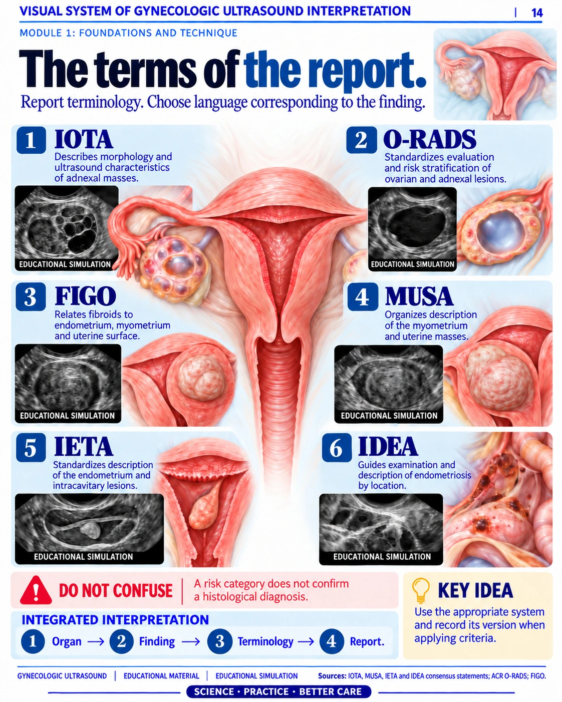
Identify the organ or structure you are describing: myometrium, endometrium, ovary, or adnexal region.
Compare the purpose of IOTA, O-RADS, FIGO, MUSA, IETA, and IDEA. Each system organizes a different aspect of interpretation.
Follow the page sequence: organ, finding, terminology, and report. Choose the appropriate terms and recognize their limitations.
Explore 30 PDF pages: 10 from the Visual System, 10 from the Comparative Atlas, and 10 from the Cases. Open the book or tap a preview to see the details.
Pages taken from the PDFs. Open the book, turn the pages, and tap a page to enlarge it. Illustrations and cases are for educational purposes.
A visual sequence for studying the gynecologic examination, from anatomy to case interpretation.
Identify the uterus, endometrium, ovaries, and adnexal structures.
Connect the imaging plane, settings, and acquisition conditions.
Consider shape, contents, contours, and vascularity when relevant.
Study similarities, differences, and findings that need closer attention.
Bring the measurements and case information together in one interpretation.
Return to the cases, tables, and visual reference resources.
The core library: 350 pages to organize your study of gynecologic ultrasound.
A visual sequence for studying the uterus, endometrium, ovaries, and adnexa. Connect what you see with the technique, measurements, and examination context.
Side-by-side comparisons show what changes between patterns. Images and explanations help organize findings and distinguish their features.
Practice interpretation with simulated scenarios, images, and commentary. Analyze the clinical information, findings, and questions that guide interpretation.
Supporting educational material. It does not replace supervised training, current protocols, or the judgment of a qualified professional.
80 additional pages to support your reading, identify points to watch, and organize your review.
Look up concepts and visual references whenever a word raises a question, without losing your place in the material.
Review technical limitations, artifacts, and comparisons that help identify interpretation errors.
Visual support for reviewing how to measure, record context, and interpret the limitations of each reference.
Unsure about the next step? Use the checklists for preparation, structures, and points to watch to organize your review.
Start with the three core resources or add the four review bonuses for just US$5.00 more.
The three main resources for building a visual approach to interpretation.
No subscription
Digital materials · In EnglishStudy, compare, practice, and review with the entire library.
No subscription
For just US$5.00 moreAdd all four reference and review resources.
Get the complete library →Digital materials · In EnglishBoth options include digital materials in English. The books and devices in the mockups are illustrative representations.
What you need to know about the format, content, and both options.
A collection of digital materials for studying and reviewing pelvic anatomy, examination technique, gynecologic findings, and simulated cases. It combines illustrations, comparisons, and visual explanations.
The Essential Library includes the three main resources: Visual System, Comparative Atlas, and 50 Gynecologic Cases — Simulated and Discussed, totaling 350 pages. The Complete Library adds four 20-page bonuses: checklists, a measurements guide, an artifacts guide, and a visual dictionary. In total, it includes 7 resources and 430 pages.
The materials and website are in English. Technical abbreviations use their customary forms.
They are PDF files. The books and screens in the images represent the digital materials; no physical product is shipped.
Yes. You can open the PDFs with a compatible reader on a computer, tablet, or smartphone. For a more comfortable view of tables and full pages, use a larger screen or zoom in.
A foundation in anatomy and ultrasound is helpful. Start with the Visual System, compare findings in the Atlas, and then practice with the simulated cases and commentary.
Yes. The guided presentation, interactive book, and enlarged previews let you explore the pages, explanations, and visual approach before choosing.
The offer includes PDF study materials. It does not include a video course, certification, or official accreditation.
No. The prices are one-time payments in US dollars: US$9.90 for the Essential Library and US$14.90 for the Complete Library. Check the final price and terms at checkout before paying.
The material presents 50 simulated cases with commentary for educational purposes. It is not medical care, and its examples cannot establish a patient's diagnosis.


